
The foundation of modern aesthetic medicine rests heavily on predictable outcomes and precise anatomical knowledge. Practitioners approach upper facial rejuvenation with a dual focus: they want to relax hyperactive muscles while still maintaining natural expressive function. The tension in this field often sits right at the intersection of standardized vs individualized dosing. We see guidelines recommending strict unit counts for specific zones; clinical reality rarely fits neatly into these predetermined boxes. You have to account for underlying facial architecture and the unique way each patient’s face moves. Muscle dynamics play a crucial role here because facial muscles never operate in isolation. They pull against each other in complex webs of tension, meaning that altering one area directly impacts the opposing groups. The depressor muscles and lifting muscles maintain a delicate physiological balance that must be respected during any aesthetic intervention.
The Primary Clinical Tool
We need to discuss the primary agent used in these procedures before analyzing exact dosage science. Botox is a highly purified form of botulinum toxin type A, which functions as a neurotoxic protein that temporarily blocks the release of acetylcholine at the neuromuscular junction. This specific chemical mechanism prevents targeted muscle contraction, allowing the overlying skin to smooth out over the course of several days. Practitioners rely on Botox to soften dynamic rhytides, primarily targeting the frontalis, glabella, and orbicularis oculi complexes. Because of its predictable spread and long-standing safety profile, high-volume clinics require a steady, reliable inventory to meet continuous patient demand. When you need to replenish your clinical stock, you can order Botox vials for your practice through secure medical distribution channels. The molecular composition of Botox requires careful handling and strict cold chain maintenance to preserve its clinical efficacy. A clinic that manages its supply protocols well ensures that every Botox session delivers consistent, top-tier results for the patient.
Navigating the Topography
Precision in medical aesthetics requires an accurate physiological map. You cannot navigate the complexities of the face without strict reliance on anatomical landmarks for injection. The upper face presents a dense landscape of neurovascular bundles, overlapping fascial planes, and interconnected muscle fibers. One slight deviation in needle placement or angle can result in undesirable outcomes like brow ptosis or an unnatural lateral arch. We rely on specific geographical markers to guide every single treatment safely.
A practitioner must confidently identify the orbital rim and supraorbital notch; these bony structures help locate the exact origin of the corrugator supercilii. They also warn the injector to avoid the critical neurovascular pathways exiting the skull in this region. If a practitioner places a bolus too close to the orbital rim, the risk of the neuromodulator migrating downward into the levator palpebrae superioris increases sharply. This specific product migration remains the primary cause of a medically induced droopy eyelid.
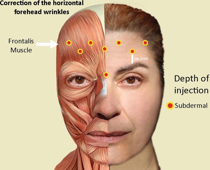
Another critical vertical marker is the midpupillary line. This imaginary vertical axis acts as a strict boundary for lateral frontalis injections. If you ignore this boundary and fail to treat the area laterally when necessary, you risk leaving the outer fibers of the frontalis highly active while the medial fibers remain paralyzed. The end result is a highly unnatural peaking of the brow that looks distinctly artificial.
We must also evaluate the hairline and brow position before drawing up a single syringe. The resting distance between the brow and the hairline dictates the available vertical space for product distribution. A narrow forehead with a low hairline requires a completely different approach than a high, expansive forehead with heavy, low-set brows. A practitioner treating a low-set brow too heavily in the lower frontalis will almost certainly cause the brow to drop further, creating a heavy and fatigued appearance for the patient.
Observing the Machinery in Motion
Anatomy gives you the static map; you still need to observe the machinery in motion. This diagnostic phase requires a thorough muscle strength assessment (rest vs animation) during the initial consultation. You ask the patient to frown aggressively, raise their eyebrows as high as possible, and smile tightly. This active process reveals the true contractile force of the target muscles. Some patients possess hyperkinetic muscles that recruit neighboring resting fibers during animation. Others show strong resting tone with surprisingly minimal dynamic movement.
We also have to account for inherent biological variables that alter drug efficacy. A 2020 clinical review published in cosmetic dermatology literature found that male patients typically require significantly higher doses of neuromodulators due to greater baseline muscle density. Sex and muscle mass differences in dosing dictate that a standard protocol designed for a female glabella will often fail to achieve full temporary paralysis in a male patient. The corrugator muscles in men are usually much thicker and extend further laterally toward the temporal fusion line. You have to adjust the units per treatment area accordingly to match the physical mass of the muscle you are actively trying to inhibit.
The Science of Measurement
The aesthetic industry frequently pushes for dosing standardization to ensure overarching safety and reproducible outcomes across different clinics. Established neuromodulator treatment protocols offer an excellent baseline for new practitioners entering the field. They provide safe injection zones and conservative unit counts to prevent major complications during the initial learning curve. Fixed-dose vs patient-specific treatment plans remain a heavily debated topic at international medical conferences. Strict adherence to a universal unit count often ignores the physiological reality of the unique patient sitting in the treatment chair. The superior alternative involves adapting the standardized baseline to fit the specific architecture of the individual face.
You also have to control the exact physical properties of the product before it ever enters the skin. Careful reconstitution and dilution practices ensure you know exactly how many active units are contained in every 0.1 mL of fluid. A standard dilution ratio of 2.5 mL of bacteriostatic saline per 100-unit vial remains common practice worldwide. Some advanced practitioners adjust this fluid ratio based on the desired physical spread of the product; a higher dilution might be used to encourage wider diffusion across the broad, thin frontalis muscle.
A practitioner must actively monitor three main delivery variables during the actual procedure:
- The exact volume of fluid administered per injection site to prevent unwanted spread.
- The angle of the needle upon entering the skin to target specific muscle bellies.
- The distinct physical resistance felt as the needle passes through different fascial planes.
The actual mechanical delivery mechanism is just as crucial as the dilution ratio. You must actively choose the correct injection depth (intramuscular vs intradermal) for each distinct anatomical zone. The glabella typically requires deep intramuscular injections to reach the bulky, deep-seated corrugators effectively. The frontalis often benefits from more superficial intradermal or subdermal placement. Superficial placement in the forehead helps the injector avoid complete diffusion into the deeper muscle belly, maintaining some baseline animation and preventing a frozen, unnatural appearance.
Archiving the Clinical Data
All of this detailed physiological assessment means very little if you do not record it properly. Meticulous treatment documentation and charting separate the amateur from the dedicated medical professional. You need a permanent record of exact units used, specific dilution ratios, and precise anatomical placement mapped out for every single session. When the patient returns in three or four months for a maintenance dose, that clinical chart serves as the only reliable way to adjust the dose safely or reproduce an exceptionally great result. A well-maintained chart acts as a clinical blueprint for future success.
The timeline for clinical review carries significant weight in the overall success of the procedure. Clear communication regarding touch-up and follow-up timing prevents unnecessary anxiety for the patient. Maximum chemical effect takes up to two full weeks to materialize completely at the neuromuscular junction. A mandatory review scheduled exactly at the 14-day mark allows you to assess the final stabilized outcome objectively. You can then add minor micro-adjustments without the risk of stacking doses prematurely.
